Genetics Talk
Published on 04/05/16
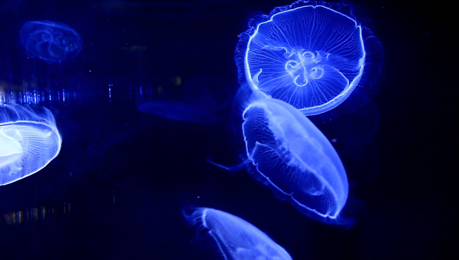 Thanks to the organisation of the science society, Rhys Jones gave a fascinating talk to a few sixth form students on the ability of scientists to add a fluorescent protein to proteins of biological interest in order to follow its progress through organisms. This technique is used by scientists who don't have access to more expensive Scanning Electron Microscopes to see structures inside cells. It basically consists of cutting a section of a protein that creates the fluorescent glow of jellyfish and pasting it onto the end of the protein that the scientist wants to observe. This can then be easily seen by scientists as it goes through synthesis, transport through the cell, and different functions.
The students were also able to see cutting edge images of 3D constructions of cell structures, learning that chloroplasts are actually long, spread out structures rather than the dome-like structures that we see in text books. The make up of these images were obtained by placing many photos of different layers and angles of the organelle together, with the final product consisting of an incredibly accurate reconstruction that can even show pathways through the organelle, whose function is still unknown. This extension from our normal biology lessons was extremely interesting and showed us how lesson work is relative to the discoveries of current biological scientists.
The students are extremely grateful to Rhys Jones for taking time out to speak about his work, and we hope to hear about any more developments in his research.